
I.A.3 Back: Spinal Cord, Meninges, CSF, Typical Spinal Nerve: Page 1 of 4
Objectives:1.3.1 Identify the spinal cord, conus medullaris, and the cauda equina. Identify the major coverings (meninges) of the spinal cord. Indicate the relationship of the meninges to the vertebral canal, cerebral spinal fluid, nerve roots and spinal cord. 1.3.2 Identify the components of a typical spinal nerve including sensory and motor portions, ventral rami, dorsal root ganglia, and demonstrate the parts of a typical spinal nerve that contain components of the visceral nervous system. |
Assigned Readings:Moore's COA pages: 128-135. See blue boxes on pages 137-139 for clinical correlations. See the yellow box on page 139 for "The Bottom Line." |
 |
| 🔍 |
| Step 1: Begin with a view of the sagittal plane. | |

|
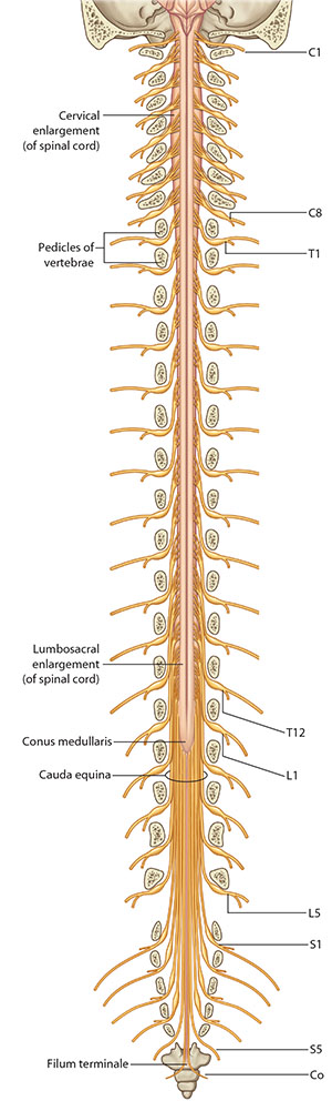
Step 2: Using the figure of the spinal column, discuss the spinal nerves with respect to their origin from the vertebral column. |
 |
How many cervical vertebrae are there? |
|
Seven. |
|
|
How many cervical nerves are there? |
|
Eight. |
|
|
Why is there an discrepancy between the number of vertebrae and the number of nerves? |
|
For the first 7 vertebrae the nerves exit cephlad (superior) to the vertebrae. From thoracic vertebra TI through the remainder of the spinal column, the nerves exit inferiorly. Cervical nerve CVIII exits inferior to cervical vertebrae CVII and allows for the pattern of one skeletal nerve per intervertebral space. |
|

|
Step 3: Using the Highlight tool, locate the spinal cord, conus medullaris, and cauda equina. |